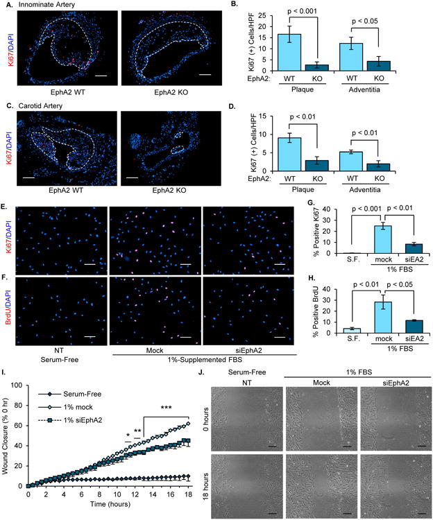
Figure 6. EphA2 deficiency attenuates proliferation in atherosclerotic lesions and hCoASMCs in vitro.

A-D) EphA2 WT and EphA2 KO mice were fed Western diet for 16 weeks, and proliferation in the (A/B) innominate and (C/D) carotid arteries were assessed by staining for Ki67 (red). The number of Ki67-positive cells in the plaque or adventitia was assessed, and plaque borders are indicated with a white dotted line. Scale bar = 100 μm. n = 6-7. E-H) hCoASMCs treated with or without EphA2 siRNA were maintained in either serum-free or 1% serum-containing conditions overnight, and proliferation was assessed by (E/G) staining for Ki67 (red) or (F/H) treatment with BrdU (10 μM) for 2 hours and staining for BrdU (red). The percentage of proliferating cells was determined for each condition. n = 4. Scale bar = 100 μm. I/J) hCoASMCs were plated onto collagen I (40 μg/mL) and a scratch was created. Wound closure was monitored by time-lapse imaging (3 regions per wound) for 18 hours. Wound closure was calculated as percent closure from t=0. J) Representative images from scratch wound assay. Scale bar = 100 μm. n = 4. Data are expressed as mean ± SEM. Statistical comparisons were made using Student's T-test (B, D, G, H) or One-way repeated measures ANOVA (I) with Bonferroni post-test. * p < 0.05, ** p < 0.01, *** p < 0.001 comparing EphA2 siRNA to Mock control.
