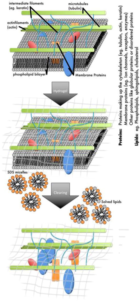
Figure 1.

Schematic diagram of the CLARITY protocol. The tissue sample is immersed in a hydrogel that binds to biomolecules with an active NH2 group such as proteins or amino acids. After polymerization of the acrylamide hydrogel within the tissue the sample is cleared using SDS detergent to wash out all tissue components that do not bind to the hydrogel such as the lipids but also other elements like iron and calcium.
