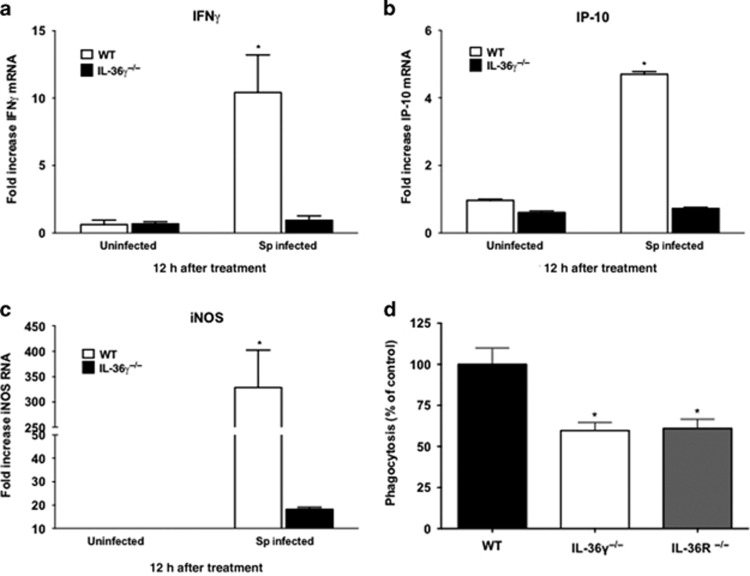
Figure 6.
Effect of interleukin-36γ (IL-36γ) on ex vivo macrophage expression of activation state markers. (a–c) Wild-type (WT) and IL-36γ−/− mice were treated with intrathecal (i.t.) Streptococcus pneumoniae (Sp) or vehicle. At 12 h after infection, interstitial and alveolar macrophages were isolated. Cytokine induction was measured by reverse transcription-PCR (RT-PCR) (*P<0.0001 as compared with uninfected WT by one-way analysis of variance (ANOVA) with Dunnett’s multiple comparisons test, n=3 per group, representative of two experiments). (d) Pulmonary macrophages from WT, IL-36γ−/−, and IL-36R−/− mice were isolated and incubated in vitro with FITC-labeled Sp (multiplicity of infection (MOI) 10:1). Intracellular fluorescence was measured at 2 h after stimulation. Phagocytosis is expressed as a percent of total fluorescence as compared with WT animals. (*P<0.01 as compared with WT by one-way ANOVA with Dunnett’s multiple comparisons test, n=6 per group, representative of two experiments). IFN, interferon; iNOS, inducible nitric oxide synthase.

