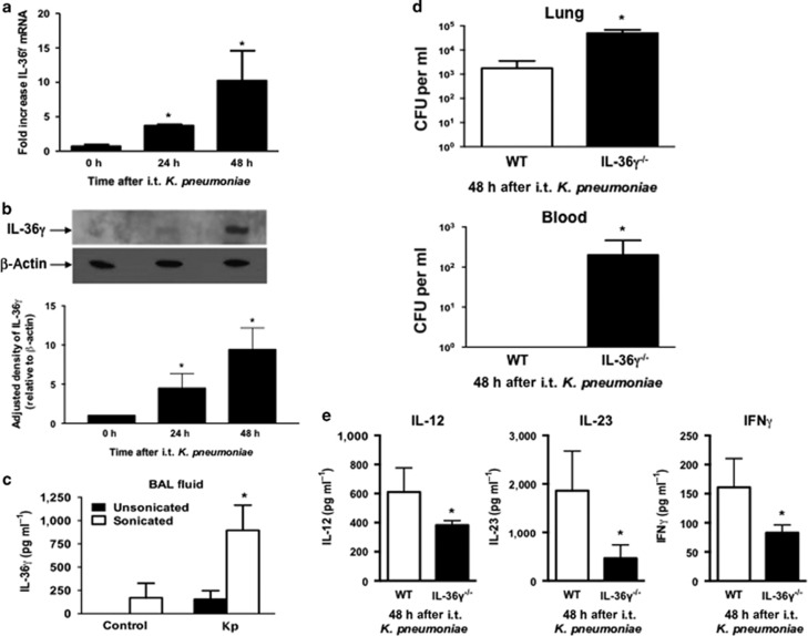
Figure 9.
Effect of interleukin-36γ (IL-36γ) on innate immune response to K. pneumonia (Kp). Wild-type (WT) mice were infected with intrathecal (i.t.) Kp and lungs were harvested at the specified time points. (a) mRNA was assessed by reverse transcription-PCR (RT-PCR) (*P<0.05 compared with uninfected controls by one-way analysis of variance (ANOVA) with Dunnett’s multiple comparisons test, n=4 mice per group, representative of three experiments). (b) IL-36γ protein was detected by western immunoblotting at 48 h after infection. Blot is representative of three mice per time point, and of two separate experiments. Densitometry represents relative density compared with β-actin from all replicates pooled from both experiments (*P<0.05 compared with uninfected mice by one-way ANOVA with Dunnett’s multiple comparisons test). (c) WT mice infected with Kp underwent BAL at 48 h after infection. BAL fluid was analyzed for IL-36γ protein by ELISA (enzyme-linked immunosorbent assay) either with or without sonication. (*P<0.05 compared with unsonicated control by one-way ANOVA with Dunnett’s multiple comparisons test, n=4–5 mice per group, representative of two experiments). (d) Lung and spleen colony-forming units (CFUs) were assessed by serial dilution at 48 h after infection (*P<0.05 compared with WT by Student’s t-test, n=8 mice per group, combined from two experiments). (e) BALF of WT and IL-36γ−/− mice was analyzed for cytokine concentration by ELISA 48 h after infection (*P<0.05 compared with WT by Student’s t-test, n=4 mice per group, representative of two experiments). IFN, interferon.

