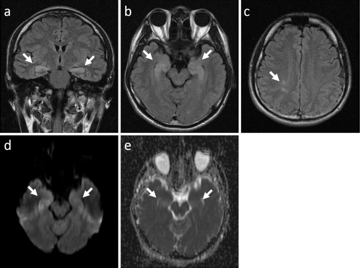
Figure 1.
Brain magnetic resonance imaging showed high-intensity areas in the bilateral hippocampi (a and b, arrows) and periventricular white matter (c, arrow). Diffusion-weighted imaging (d) and apparent diffusion coefficient mapping (e) showed high-intensity signals (arrows), which were suggestive of vasogenic edema.

