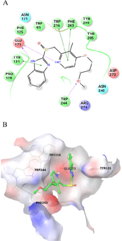
Fig. 4.

Rabeprazole binding interactions are shown in A) Ligplot. The important interacting residues are depicted using SiteMap20 and B) Crystal structure binding pocket of Arthrobacter protophormiae ENGase. Rabeprazole is shown in green sticks. The important interacting residues are shown in grey lines.
