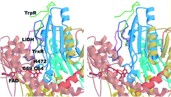
Figure 4.
Stereo view of the C-terminal extensions in enzymes of the pyridine nucleotide–disulfide oxidoreductase family. The structure of rat TrxR is shown as a ribbon model (color coding as in Fig. 3). The C-terminal extensions are shown as Cα traces (LiDH, dihydrolipoamide dehydrogenase, magenta; TrpR, trypanothione reductase, green; TrxR, rat TrxR, blue). The positions of the catalytic disulfide (Cys-59–Cys-64), and the cysteine residues Cys-497 and Cys-498 in the C-terminal tail of TrxR are indicated by yellow spheres.

