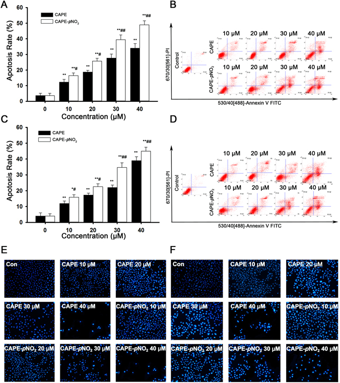
Figure 2.

CAPE and CAPE-pNO2 induce apoptosis in colon cancer cells. HT-29 and HCT-116 cells were treated with 0, 10, 20, 30 and 40 μmol/L CAPE and CAPE-pNO2 for 48 h. The apoptosis rates of HT-29 cells (A) and HCT-116 cells (B) were calculated by SigmaPlot 12.5. Flow cytometry analysis in HT-29 (C) and HCT-116 (D) cells. HT-29 cells (E) and HCT-116 (F) cells (×200) were stained by Hoechst 33342 after treatment with different concentrations of CAPE and CAPE-pNO2 for 48 h. From the microscopic vision fields, the cell number was decreased, and fluorescence was increased by treatment for 48 h in a dose-dependent manner. Values represented the means ± SD from three independent experiments, and error bars represented the STDEV (SD). *p < 0.05, **p < 0.01: CAPE and CAPE-pNO2 compared with the control. # p < 0.05, ## p < 0.01: CAPE-pNO2 compared with CAPE at the same concentration.
