
Figure 4.
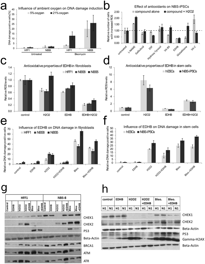
Response of NBS cells to oxidative stress and antioxidants. (a) The abundance of DNA damage measured by FACS-based detection of the DNA double-strand marker gamma-H2AX in HFF1 and NBS8 fibroblasts. DNA damage was induced by 30 µg/ml Bleomycin and compared under ambient (21%) and physiological (5%) oxygen concentrations. (b) NBS8-iPSCs were treated with either H2O2, several compounds known to influence DNA repair and ROS levels, or both. Internal ROS levels were then measured by FACS-based detection of the fluorescent ROS marker DCF-DA. The results were normalized to the untreated or peroxide-alone treated conditions respectively. (c) The influence of EDHB on internal ROS levels was tested on control (HFF1) and patient fibroblasts (NBS5, NBS8). The cells were either treated with EDHB alone or in combination with H2O2 to stimulate oxidative stress conditions. (d) Same experiment as in (c), but comparing control (hESCs) and patient (NBS-iPSCs) pluripotent stem cells. (e) The influence of EDHB on DNA damage (by detection of gamma-H2AX) was tested on control (HFF1) and patient fibroblasts (NBS5, NBS8). The cells were either treated with EDHB alone, in combination with H2O2 to stimulate oxidative stress conditions, or in combination with Bleomycin to stimulate mutagenic stress conditions. (f) Same experiment as in (e) but comparing control (hESCs) and patient (NBS-iPSCs) pluripotent stem cells. Bars indicate SD between independent experiments (n = 3). (g,h) Influence of DNA damage and EDHB on phosphorylation of DNA damage signaling proteins. Cells were treated with EDHB (antioxidant and inducer of HIFpathway), hydrogen peroxide (H2O2) and radiomimetic bleomycin (Bleo). (g) Immunofluorescent detection of phosphorylated signaling proteins in fibroblasts (HFF1, NBS-8) after SDS-PAGE. (h) Immunofluorescent detection of phosphorylated signaling proteins in hESCs (H1) and NBS-8-iPSCs (N1) after SDS-PAGE. Each lane of b-Actin corresponds to the lanes directly above and b-Actin is always unphosphorylated. For the sake of better readability western blots were cropped.