
Figure 1.
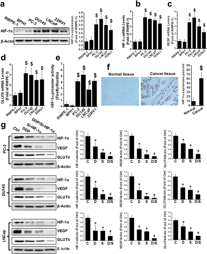
Upregulation of HIF-1α in human PCa. (a) HIF-1α protein was detected by western blot in nonmalignant (RWPE-1 and BPH1) and PCa cell lines (PC-3, DU145, LNCaP, and 22RV1) as indicated. (b–d) Total RNA extracted from RWPE-1, BPH1, PC-3, DU145, LNCaP, and 22RV1 cells was subjected to qRT-PCR for HIF-1α (b), VEGF (c) and GLUT4 (d). (e) The HIF-1α promoter-driven reporter (firefly luciferase) and a control vector (Renilla luciferase) were co-transfected into RWPE-1, BPH1, PC-3, DU145, LNCaP, and 22RV1 cells for measurement of luciferase activity. HIF-1α promoter activity was calculated as a ratio of firefly to Renilla activity. (f) Human normal and malignant tissue (Gleason score 9) sections were probed with HIF-1α antibody (scale bars, 100 µm). (g) Protein expression of HIF-1α, VEGF, and GLUT4 were examined with western blot, in PC-3, DU145, and LNCaP cells after various treatments as indicated. Data are expressed as mean ± SD of seven independent experiments. $p < 0.05 versus RWPE-1 or BPH1 cells or normal tissue. *p < 0.05 versus control group. #p < 0.05 versus si-HIF-1α or DDP group. Original blots are shown in Supplementary Figure 5. C: Ctrl; D: DDP; S: si-HIF-1α; D/S: DDP/si-HIF-1α.