
Figure 4.
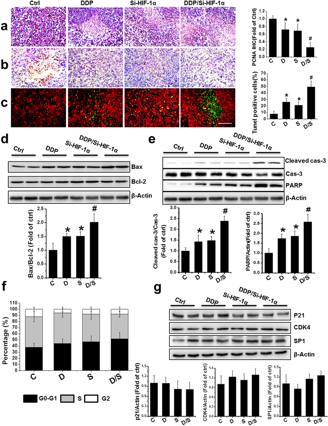
Effect of treatment with DDP and/or HIF-1α siRNA on the induction of apoptosis in PCa xenografts. (a–c) Representative images of H&E (a), PCNA (b), and TUNEL (c) in the xenografts with treatments as indicated (scale bars, 100 µm). (d,e) Bax/Bcl-2 ratio (d) and protein abundance of cleaved forms of caspase-3 and PARP (e) were examined in PCa xenografts by western blot. (f) Values represent the percentage of cells in each phase of the cell cycle that were detected by flow cytometry analysis. (g) Western blot for protein expression of p21, CDK4, and SP1 after various treatments. Data were presented as mean ± SD (n = 6). *p < 0.05 versus control group; #p < 0.05 versus si-HIF-1α or DDP group. The original blots are presented in Supplementary Figure 7.