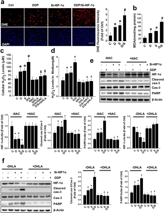
Figure 5.

ROS overproduction mediated apoptosis induced by combined treatment of DDP and HIF-1α in PC-3 xenografts and cell culture. (a) ROS monitored by DHE (red) and nuclei by DAPI (blue) staining in PCa xenografts (scale bars, 50 µm). (b) MDA formation of PCa xenografts was examined after various treatments (c,d). PC-3 cells were treated with DDP, si-HIF-1α plasmid, or both, in the presence or absence of NAC (5 mM) or DHLA (0.25 mM) for 24 h. Total lysates (c) and culture media (d) were used to detect cellular H2O2 level. (e,f) Western analysis for HIF-1α as well as cleaved caspase-3 and PARP in PC-3 cells following various treatments. Data were presented as mean ± SD of three independent experiments. *p < 0.05 versus control group; #p < 0.05 versus si-HIF-1α or DDP group; $p < 0.05 versus DDP/si-HIF-1α group. The original blots are presented in Supplementary Figure 8.
