Figure 1.

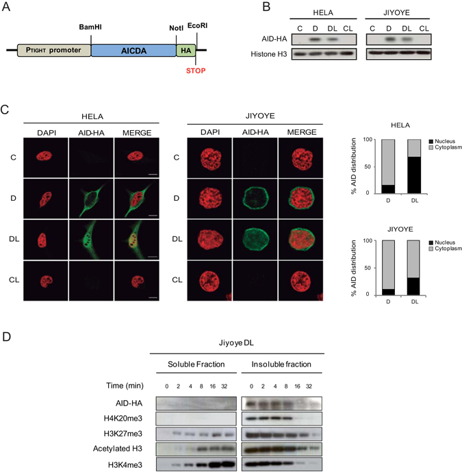
An inducible system for AID expression and accumulation in the nucleus. (A) Representative scheme of the HA-tagged AID retroviral construct. (B) AID expression is induced following doxycycline (D) treatment and retained in the nucleus upon additional treatment with leptomycin B (L), which specifically inhibits nuclear export. Anti-HA antibody was used to detect AID expression and histone H3 was used as a loading control (C) Representative confocal images of AID subcellular localisation in HeLa and Jiyoye cells after doxycycline and leptomycin B treatment. Nuclear DNA was counterstained with DAPI (red). A total of 25 cells from randomly selected fields were analysed in each experimental condition. The graphs in the right show the quantification of the cellular signal of AID within the cells. Light gray section of the bar indicates the average percentage of cytoplasmic AID signal. Black section of the bar indicates the average percentage of nuclear AID signal. (D) Association of AID with heterochromatin. Time course digestion of HA-tagged AID Jiyoye cells nuclei with DNase I, in which the supernatant contains the euchromatin fraction (as demonstrated by the appearance of H3Ac and H3K4me3) and the pellet contains the heterochromatin fraction (as shown by the progressive decrease of H4K20me3 and H3K27me3).
