
Figure 6.
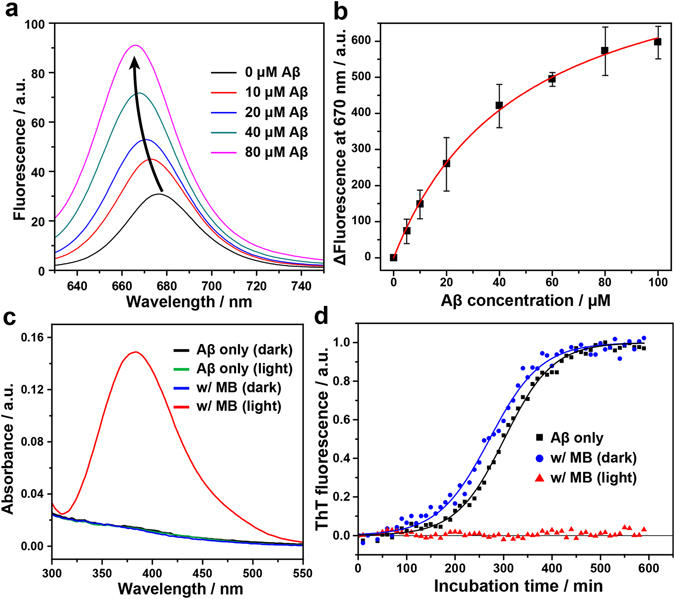
Photo-excited MB inhibits the Aβ42 aggregation by means of the binding affinity of MB and the photo-oxidation of Aβ42. (a) Change in fluorescence spectra of 0.2 μM MB upon addition of various concentrations of the Aβ42 monomer. (b) Fluorescence binding affinity assay between Aβ42 and MB. The fluorescence of MB (0.2 μM) at 670 nm was measured with increasing concentration of Aβ42 from 0 to 100 μM. Binding constant Kd was derived from the fitted curve. (c) DNPH assay to monitor a carbonyl content in the Aβ42 peptide. DNPH reacts with the carbonyl groups in oxidized peptides, resulting in the formation of a DNP hydrazone product, which shows an absorption maximum of near 380 nm. (d) The kinetics of Aβ42 fibril formation monitored at 30 °C by ThT fluorescence in the absence and presence of MB under dark or light conditions. For the light condition, the MB-treated Aβ42 samples were irradiated with LED light for 30 min at 4 °C before the measurement. Each point is an average of the fluorescence signal of at least four wells containing the same solutions. Lines indicate fits of a sigmoidal growth curve.