
Figure 1.
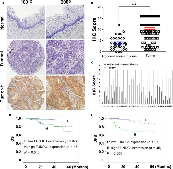
FUNDC1 expression in cervical cancer tissues and its prognostic significance in patients. (A) Normal: A correspondence normal cervical tissue (Case 12) shows negative expression of FUNDC1 (IHC score: 0). Tumor‐L: A cervical cancer tissue (Case 37) exhibited low expression of FUNDC1 (IHC score: 2). Tumor‐H: A cervical cancer tissue (Case 5) exhibited high expression of FUNDC1 (IHC score: 12). (B) Statistical analysis revealed significantly higher expression of FUNDC1 in cervical cancer tissues (**P < 0.01, Student's t‐test). (C) The expression of FUNDC1 in 35 cervical cancer samples which have paired adjacent normal lung tissues. (D and E) Kaplan–Meier plots showed overall survival (D) and disease‐free survival (E) curves of our enrolled 82 patients, according to FUNDC1 expression levels in the primary tumor (L, low expression of FUNDC1; H, high expression of FUNDC1. Log‐rank test).