
Figure 4.
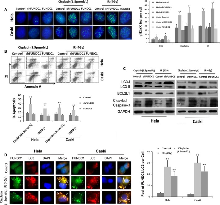
Depletion of FUNDC1 promotes cisplatin and IR‐induced DNA damage and apoptosis of cervical cancer cells. (A) Silencing of FUNDC1 inhibits the repair of cisplatin and IR‐induced DNA damage. Cells were subjected to cisplatin and IR at indicated dose, 12 h later, fixed for immunofluorescence. Shown is staining with antibodies to γH2AX (green). γH2AX foci used as a measure of unrepaired DNA damage. (B) Knockdown of FUNDC1‐enhanced Cisplatin and IR‐induced cell apoptotic events in Hela229 and CaSKi cells monitored by Annexin V‐APC/PI staining and flow cytometry assays (the rate of cell apoptosis = D2% + D4%). (C) After treatment with Cisplatin or IR, LC3, BCL2L1, and cleaved Caspase‐3 were determined by Western blotting, and GAPDH was used as a normalized control. (D) Cells treated with IR or Cisplatin were visualized by immunofluorescence with anti‐FUNDC1 (green) and anti‐LC3 (red). Merged foci showed interaction between FUNDC1 and LC3. The average numbers of IR and Cisplatin‐induced foci of FUNDC1/LC3 per cell were quantified with Image J software. All data represent the mean ± SE derived from three individual experiments with triplicate wells. (*P < 0.05, **P < 0.01, Student's t‐test)