
Figure 3.
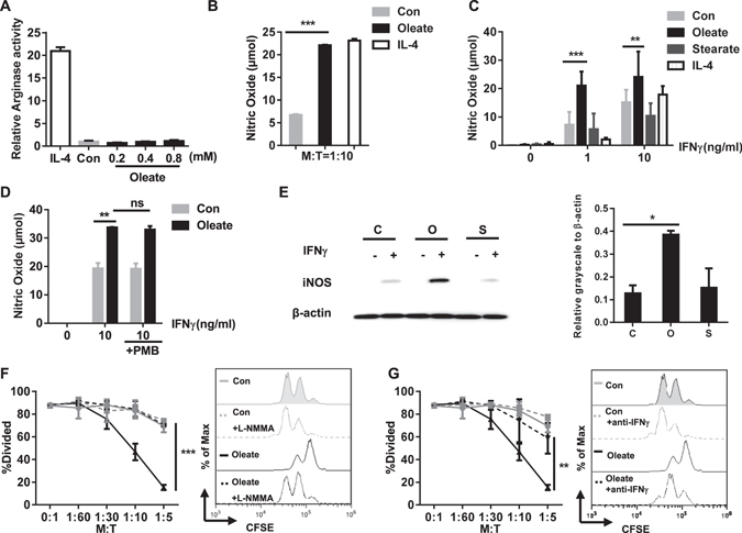
The sodium oleate-induced regulatory phenotype of MSC-2 cells depends on NO production but not arginase activity. MSC 2 cells were cultured in the presence of either IL-4 (100 ng/ml), BSA (Con) or sodium oleate (0.2 mM). After 24 h arginase activity was determined (A). MSC-2 cells were co-cultured with purified CD4+ T cells for 72 h in the presence of either BSA, sodium oleate (0.2 mM) or IL-4 (100 ng/ml) and NO was measured via Griess reagents in the supernatant (B). MSC-2 cells were stimulated with the indicated concentrations of IFNγ for 24 h in the presence of either BSA (Con), sodium oleate (0.2 mM), sodium stearate (0.2 mM), IL-4 (100 ng/ml) and IFNγ as indicated. NO was measured via Griess reagents in the supernatant (C). MSC-2 cells were treated with BSA (Con), sodium oleate (0.2 mM) in the presence or absence of IFNγ (10 ng/ml) and polymyxin B (10 µg/ml) as indicated (D). MSC-2 cells were pretreated overnight with BSA (C), sodium oleate (O) or sodium stearate (S) and subsequently stimulated for 8 h with IFNγ (1 ng/ml). Cell lysates were analyzed by Western blot analysis for iNOS as well as beta-actin expression. Quantitative evaluation in relation to β-actin expression was performed. Shown is the mean ± SD of n = 2 experiments (E). MSC-2 cells were co-cultured with CD4+ T cells in the ratios indicated and in the presence or absence of BSA (Con), sodium oleate (0.2 mM) and the iNOS inhibitor L-NMMA (2.5 mM) (F) or anti-IFNγ (10 µg/ml) (G). Cell proliferation was evaluated by CFSE staining after 72 h. Shown is the mean ± SD from two to four independent experiments. *p < 0.05; **p < 0.01; ***p < 0.001.