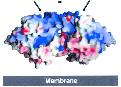
Figure 2.
Electrostatic surface potential of the CA XII dimer. The dimer twofold axis lies vertically in the center of the figure, perpendicular to the membrane surface. Arrows indicate the active site clefts, which are hidden in this view. Surface calculated and color-coded from −15 kT (red) to + 15 kT (blue) with GRASP (63).

