
Figure 1.
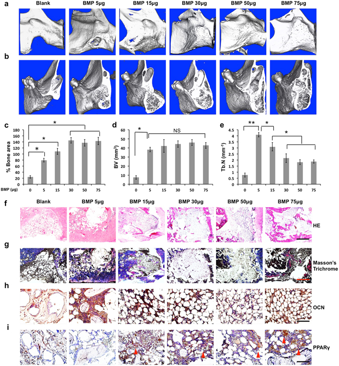
BMP2 dose ranging from 0 to 75 μg (0–1.5 mg/mL) was used to induce cyst-like void bone formation with a dose-dependent fashion in the rat critical-sized (5 × 5 mm) mandibular defect. 8 weeks postoperatively, the collected mandibular implants were measured by the following analysis: Micro-computed tomography (microCT) images in general (a) and sagittal sections (b); Quantification of % bone surface area (c), bone volume (mm3) (d), and trabecular number (Tb.N, mm−1) (e); Histological analysis including HE staining (scale bar = 500 µm) (f), Masson’s trichrome staining (scale bar = 500 µm) (g), and immunohistochemical staining for OCN (scale bar = 50 µm) (h) and PPARγ (scale bar = 50 µm) (i). Red arrowhead indicates high expression of PPARγ. Blank, blank scaffold; BMP, BMP2; NS, no significant difference. Data presented as means ± SD (n = 3/group); *p < 0.05, **p < 0.01.