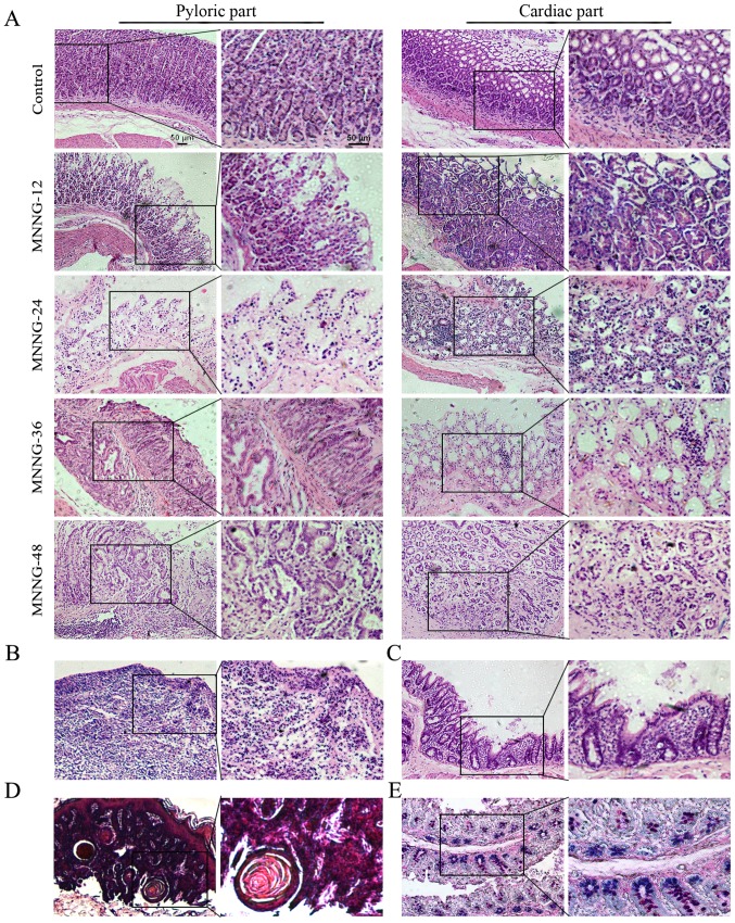
Figure 2.
Histology of the control and MNNG-induced rat gastric tissues. (A) Superficial gastritis was noted in the MNNG-12 group, atrophic gastritis in the MNNG-24 group and the cardiac part of the MNNG-36 group, and atypical hyperplasia in the pyloric part of the MNNG-36 and MNNG-48 groups. (B) H&E staining of ulcers in the MNNG-36 group. (C) H&E staining of gastric tissues in the MNNG-36 group. Intestinal metaphase changes were observed. Goblet cells were noted in the gastric mucosa. (D) H&E staining for gastric tissuse in the MNNG-48 group. Esophageal-like sarcoma lesions were observed. (E) Small intestinal type metaplasia was detected using AB-PAS staining. MNNG-12, 24, 36 and 48 represent the rats treated with MNNG for 12, 24, 36 and 48 weeks, respectively. Original magnification of the left panel, ×100; the right panel, ×200; scale bar, 50 µm.

