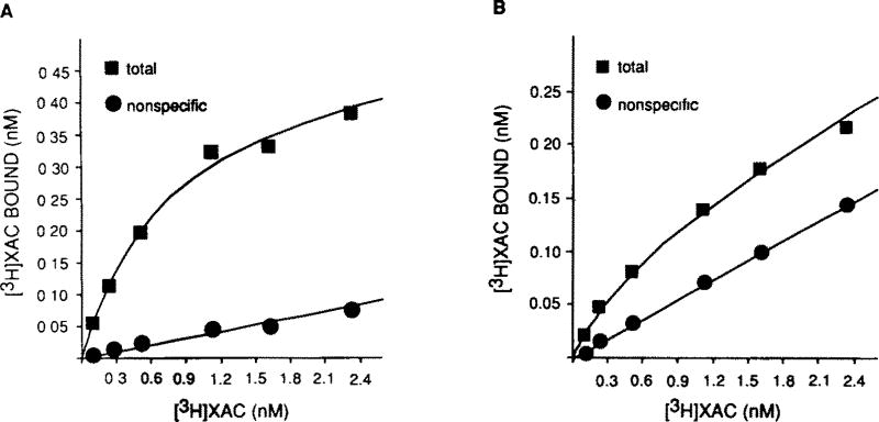
Fig.1.
[3H]XAC saturation curves in (A) solubilized bovine brain membranes and (B) XAC-Affi-Gel-purified preparation. Membrane solubilization, affinity chromatography and binding assays were performed as described in section 2. Non-specific binding was defined with 10 µM R-PIA or 5 mM theophylline. Gpp(NH)p (10 µM) was present in all assay tubes. These plots are representative of 5 similar experiments.

