Fig. 3.

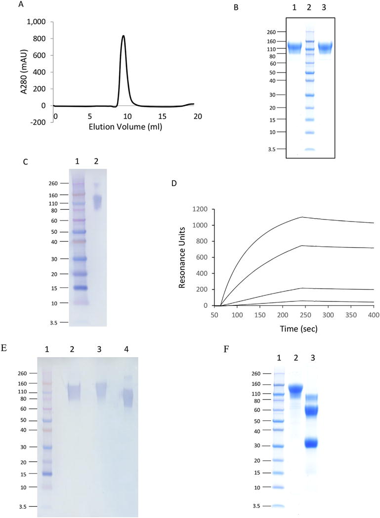
Characterization of GP1 protein purified by Ni column. A) Size exclusion chromatography elution profile. GP1 protein sample was loaded onto a Superdex 200 HR 10/30 column with PBS as a running buffer. B) SDS-PAGE analysis. GP1 samples were loaded onto a 4–12% Bis-Tris protein gel and stained with ProBlue. Lane 1, nonreducing conditions; Lane 2, protein markers; Lane 3, reducing conditions. C) Western blot analysis. 0.5 μg of purified GP1 was electrophoresed on a 4–12% SDS-polyacrylamide gel, transferred by electroblotting to a PVDF membrane, and visualized by immunostaining as described in the text. Under nonreducing conditions: Lane 1, protein markers; Lane 2, purified GP1. D) BIAcore binding sensorgram between GP1 and mouse anti-Zaire GP monoclonal antibody. The antibody was immobilized on a CM5 chip and GP1 served as an analyte. The concentrations of GP1 was 40, 5, 0.625 and 0.078 μg/ml respectively. E) Western blot analysis of GP1 deglycosylation. Under reducing conditions: Lane 1, protein markers; Lane 2, neuraminidase treated GP1; Lane 3, untreated GP1; Lane 4, PNGase F treated GP1. F) SDS-PAGE analysis of GP1 (Lane 2) and GPΔMuc (Lane 3) under reducing conditions. Lane 1, protein markers.
