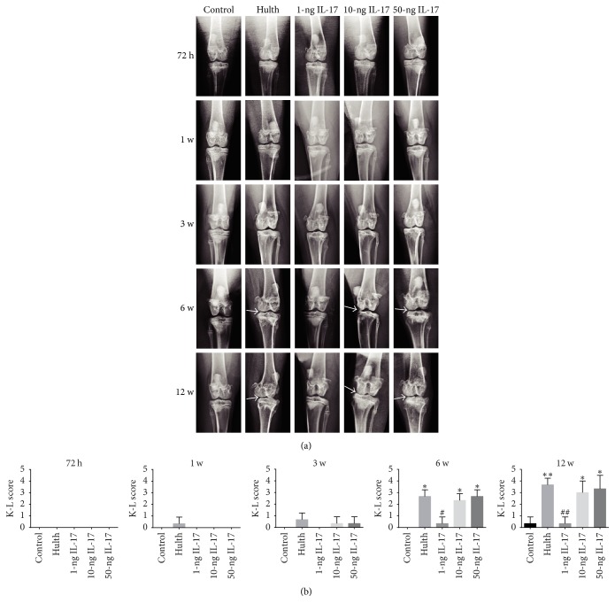
Figure 2.
(a) Radiological evaluation shows that the control group exhibited normal knee joint signs at all 5 time points. Similar to the control group, the 4 experimental groups displayed no or mild signs of degeneration at 72 h, 1 week, and 3 weeks. At 6 weeks and 12 weeks, severe radiological signs of osteoarthritis were observed in the Hulth group, the 10-ng group, and the 50-ng group, while only a slight narrowing of the joint space and little osteophyte formation were seen in the 1-ng group. Arrows indicate narrowing of the joint space and osteophyte formation. (b) Kellgren-Lawrence scores (K-L score) for the severity of osteoarthritic lesions. ∗Compared to the control group, P < 0.05, ∗∗P < 0.01. #Comparing the 3 IL-17 groups to the Hulth group, <0.05, ##P < 0.01.

