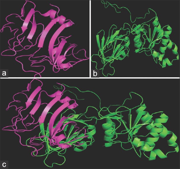
Figure 2.

Structural comparison of Staphylococcus aureus NAD kinase (magenta) and human NAD kinase (Green) using PyMol. (a) Staphylococcus aureus NAD kinase; (b) human NAD kinase; (c) super imposed structures of human NAD kinase (green), staph NAD kinase (magenta)
