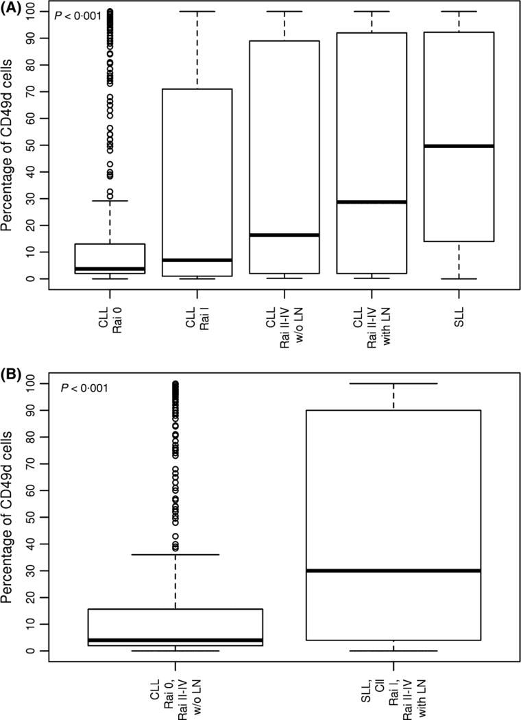
Fig 1.
Association between CD49d expression at time of diagnosis and type of CLL presentation. The percentage of lymphocytes expressing CD49d is shown on the y-axis (each bar shows median, first, and third quartiles). (A) By Rai stage; (B) By lymphadenopathy. CLL, chronic lymphocytic leukaemia; LN, lymphadenopathy; SLL, small lymphocytic leukaemia; w/o, without.

