Figure 1.

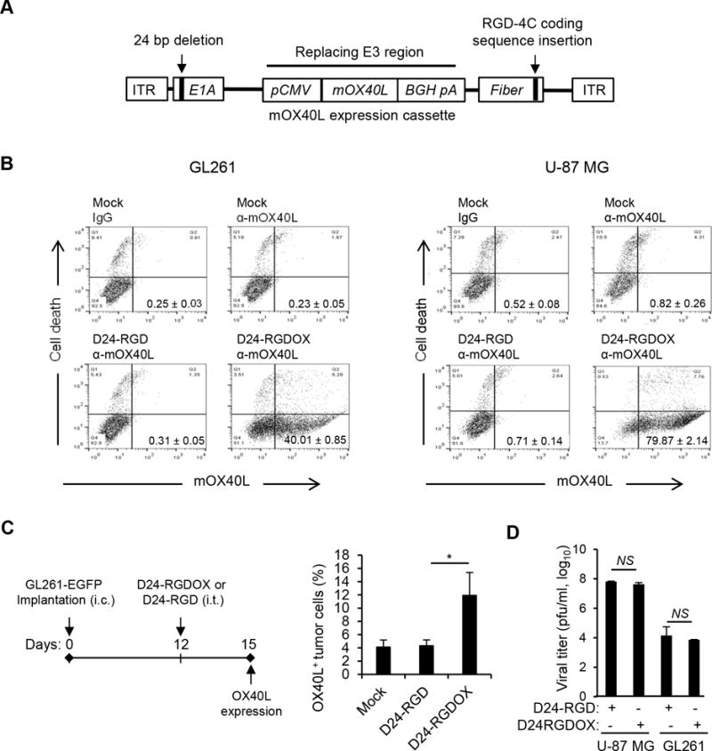
Construction and characterization of Delta-24-RGDOX. A, Schematic representation of the Delta-24-RGDOX genome, including a 24-base pair deletion in the E1A gene that encodes an RB-binding region and an insertion in the fiber gene that encodes an integrin-binding motif (RGD-4C) in the HI loop of the protein. The mouse OX40L (mOX40L) expression cassette, including the cytomegalovirus (CMV) promoter (pCMV), mOX40L cDNA, bovine growth hormone poly-adenylation signal (BGH pA), replaces the E3 region of the human Adenovirus 5 genome. ITR: inverted terminal repeat. B, Expression of mOXO40L by Delta-24-RGDOX in mouse GL261 and human U-87 MG glioma cells. Cells were infected with Delta-24-RGD or Delta-24-RGDOX at 100 (GL261) or 10 (U-87 MG) PFU/cell. After 48 hours, the cells were harvested, and mOX40L expression and cell death (cells with broken membrane stained with ethidium homodimer-1) were analyzed with flow cytometry. Representative dot plots for each analysis are shown. The numbers at the lower right corners indicate the percentages of live cells expressing mOX40L on their cell membrane. C, (left panel) A cartoon depiction of the treatment scheme. i.c.: intracranial; i.t.: intratumoral. (right panel). Expression of mOX40L on tumor cells from virus-treated tumors. The hemispheres with tumors from treated mice (3 mice per group) were harvested, and the cells were dissociated and stained with anti-mOX40L-APC. The stained cells were analyzed using flow cytometry. Tumor cells were gated for EGFP+. The results from two independent experiments are shown. D, Replication of Delta-24-RGDOX or Delta24-RGD in U-87 MG and GL261 cells. Values represent the mean ± standard deviation (n = 3). * P = 0.02; NS: not significant (P ≥ 0.05), 2-tailed Student’s t test. Mock: PBS; D24-RGD: Delta-24-RGD; D24-RGDOX: Delta-24-RGDOX.
