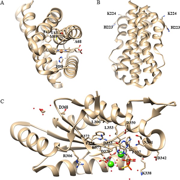
Fig 2. Positions of predicted critical amino acids in the crystal structure of EcGReg/DosC.

(A) Crystal structure of the globin domain of EcGReg/DosC (pdb: 4zvb, ferrous form) shows the locations of the critical residues in the heme pocket. H98 is the proximal histidine which binds the heme. The side chains of F42, Y43, and M69 surround the heme center. The side chain of A68 points away from the heme center. (B) Crystal structure of the middle domain of EcGReg/DosC (pdb: 4zvc, form I) shows the locations of critical residues H223 and K224, (C) Crystal structure of the DGC domain of EcGReg/DosC (pdb: 4zvf, GTPαS-bound) shows the locations of mutated residues in this work.
