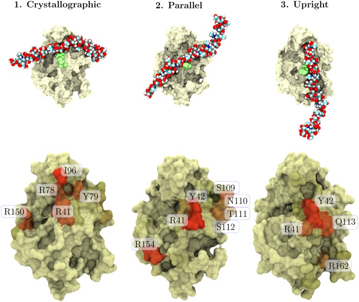
Fig 2. Molecular details of the HA–CD44 binding modes.
The three binding modes found in this study. Tan surface represents the CD44 HABD domain, the light green spheres depict R41 (important binding residue), and the multicolored rod is the HA16 oligomer. The images shown at the top are snapshots from the starting frames of the gathering simulations (Table A in S1 File). The figures at the bottom describe the most important binding residues in each binding mode. The more reddish the surface becomes, the more contacts it holds, so that red color corresponds to 350 or more contacts on average.

