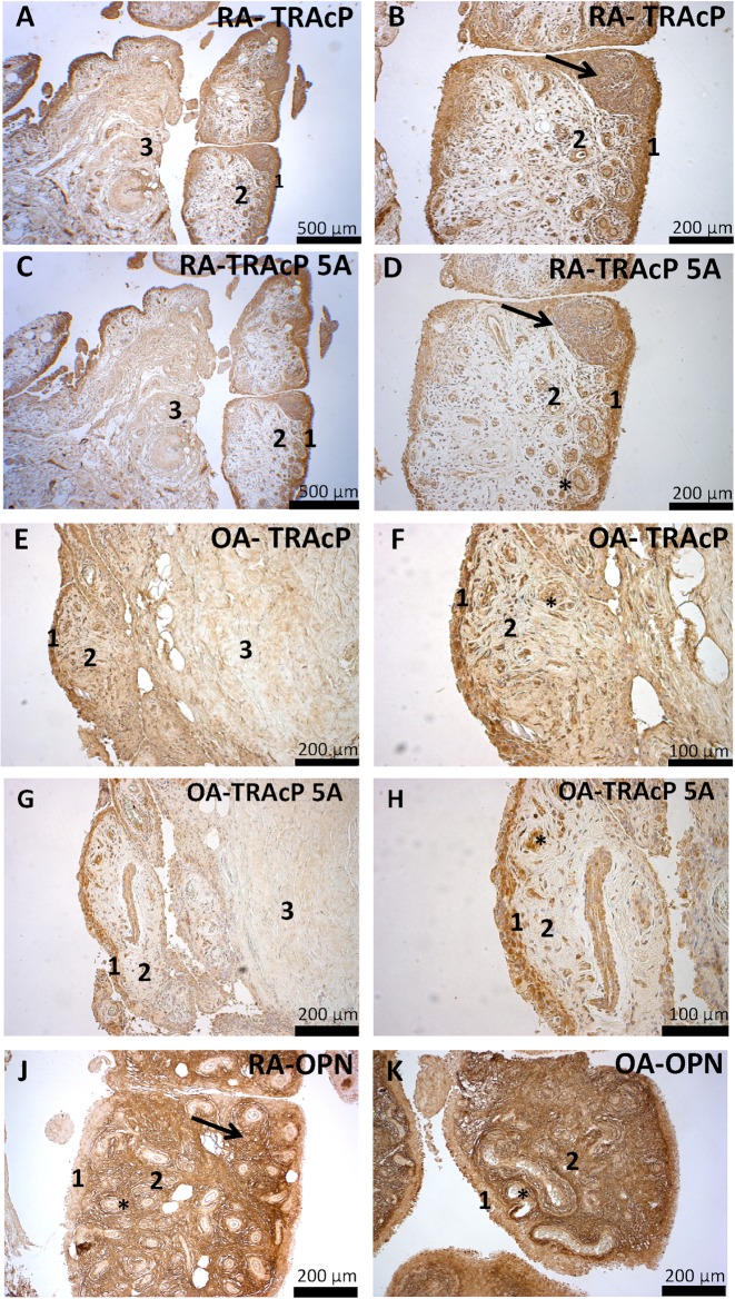
Fig 3. Synovial tissue immunohistology.
The antibody staining in each panel is shown as brown color. The generic TRAcP antibody (A, B, E, F), which stains both 5A and 5B isoforms, localized mostly in the lining layer (1) cells in a similar pattern between RA and OA, but a slightly more intensive staining of the sublining layer (2) cells and stroma could be seen in OA samples. The pattern of staining within deep stroma (3), which consists mostly of dense connective tissue and fat, was also similar between the diseases. TRAcP 5A stain (C, D, G, H) localized similarly to the generic antibody, and no differences were found between staining intensities of the sample groups. This indicates that most of TRAcP in the synovial tissue is likely in the 5A form, but differences in TRAcP 5B levels within the synovial tissue are possible between OA and RA. Both TRAcP antibodies were also localized in endothelial (asterisk) and lymphatic cells within the lymphatic follicle (arrow). OPN antibody stain (J, K) localized in the extracellular matrix, with only slight intracellular staining seen. OPN staining was most intense in the sublining layer (2), while the stainings of the lining layer (1) and deep stroma were lighter. OPN antibody did not localize in the endothelial cells (asterisk) but was found within the lymph node (arrow) stroma and some cells within it. No difference was found in the pattern or intensity of staining between RA and OA samples.

