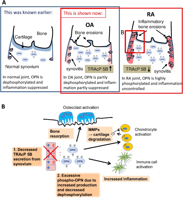
Fig 4. Schematic presentations of OPN’s role in OA and RA.
Picture A displays a schematic illustration of the normal, osteoarthritis and rheumatoid arthritis synovial cavity. The phosphorylation of OPN in synovial fluid gradually increases from healthy to RA in correlation with the severity of the inflammation and symptoms. Picture B is an illustration of possible cell level actions influenced by OPN in the pathogenesis of RA. Insufficient TRAcP 5B production in synovial tissue may lead to an excessive phospho-OPN concentration, which leads to elevated immune cell activation, more cartilage destruction and greater activation of bone resorbing osteoclasts, all effects contributing to the pathogenesis of RA.

