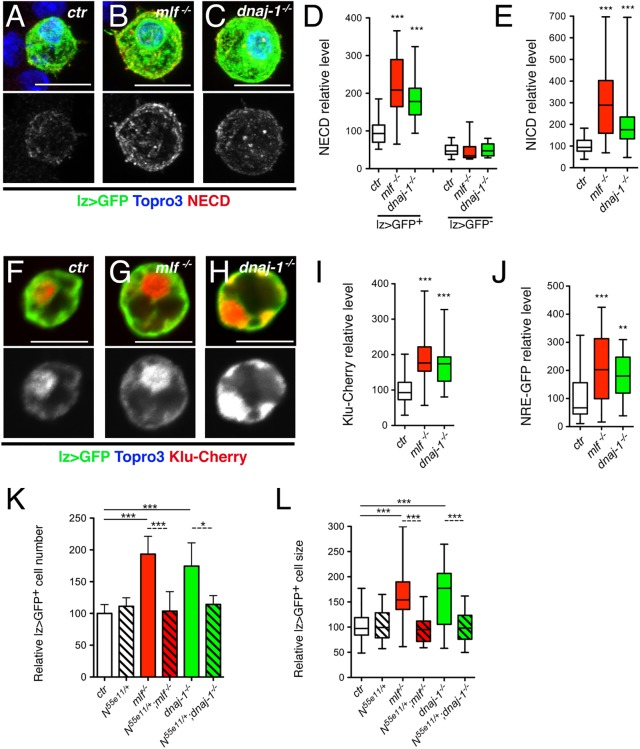
Fig 6. The increase in lz>GFP+ cell number and size in mlf and dnaj-1 mutant larvae is caused by overactivation of the Notch signaling pathway.
(A-C) Immunostainings against Notch (NECD: Notch extracellular domain) in blood cells from lz-GAL4,UAS-mCD8-GFP/+ control (A), mlf-/- (B) and dnaj-1-/- (C) larvae. The immunostaining against Notch protein only is shown in the lower panels. Nuclei were stained with Topro3. (D) Quantification of NECD immunostainings in lz>GFP+ and lz>GFP- blood cells from control, mlf-/- and dnaj-1-/- larvae. (E) Quantification of NICD (Notch intracellular domain) immunostainings in lz>GFP+ blood cells from control, mlf-/- and dnaj-1-/- larvae. (F-H) Expression of the Notch pathway reporter Klu-Cherry in lz>GFP+ blood cells from control, mlf-/- or dnaj-1-/- larvae. Klu-Cherry expression only is shown in the lower panels. (I) Corresponding quantification of Klu-Cherry level. (J) Quantification of the expression level of the Notch pathway reporter NRE-GFP in PPO1-expressing cells from control, mlf-/- or dnaj-1-/- larvae. (K, L) Relative lz>GFP+ blood cell number (K) and size (L) in third instar larvae of the indicated genotypes.

