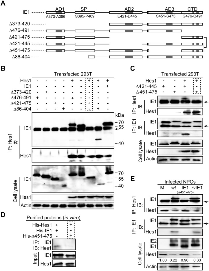
Fig 4. The IE1/Hes1 interaction requires AA451-475 of IE1.
(A) Schematic diagram of wild-type IE1 and IE1 mutants used in this study. (B-C) IE1 region required for IE1/Hes1 interaction in vivo. Following co-transfection with 6μg plasmid pCDH-Hes1 and 6μg pEYFP -based plasmids expressing wild-type or mutant IE1 (Δ373–420, Δ476–491, Δ421–475, Δ86–404, Δ421–445 and Δ451–475) for 48h, 293T cells were sequentially processed for Hes1-directed IP and IB for IE1 and Hes1. The total IE1 and Hes1 protein levels in cell lysates were also assessed. Band corresponding to IgG heavy chains are indicated by arrows. (D) Interaction of purified IE1 or Δ451–475 and Hes1 in vitro. 1μg of each purified protein was mixed and subjected to pull down assay as described in Materials and Methods. (E) Interaction between IE1 or Δ451–475 and Hes1 in infected NPCs. Following mock (M), TNwt (wt), TN-IE1(Δ451–475) (IE1(Δ451–475)) or TNrvIE1 (rvIE1) infection at an MOI of 10, NPCs were harvested at 12hpi and cell lysates were subjected to either Hes1- or IE1-directed IP analysis and subsequent IB for IE1 or Hes1, respectively. The total IE1/2 and Hes1 protein levels in cell lysates were also assessed. The band corresponding to IgG heavy chain is indicated by an arrow.

