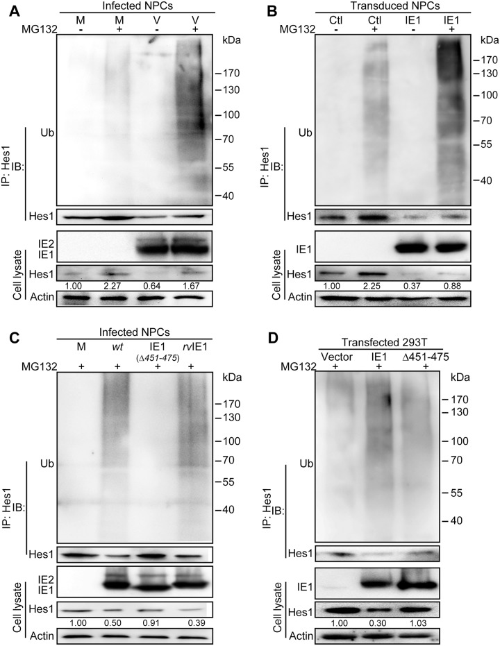
Fig 5. IE1 prompts Hes1 ubiquitination in vivo.
(A) Effect of HCMV infection on ubiquitination of endogenous Hes1 in NPCs. Following mock (M) or HCMV infection (V) for 3h (MOI = 3), NPCs were treated with 12.5μM MG132 (+) or equivalent volume of DMSO (-) for 9h, and harvested at 12 hpi. Cell lysates were subjected to Hes1-directed IP and subsequent IB for ubiquitin (Ub) and Hes1. (B) Effect of IE1 on ubiquitination of endogenous Hes1 in transduced NPCs. NPCs were transduced with lentivirus expressing IE1 (IE1) or control (Ctl). Cells were treated with MG132 (+) or equivalent volume of DMSO (-) when GFP signal was clearly observed, harvested after being treated for 12 h, and subjected to Hes1-directed IP and subsequent IB for ubiquitin (Ub) and Hes1. (C) Effect of IE1 AA451-475 on ubiquitination of endogenous Hes1 in HCMV infected NPCs. NPCs were subjected to mock (M), TNwt (wt), TN-IE1(Δ451–475) (IE1(Δ451–475)) or TNrvIE1 (rvIE1) infection at an MOI of 10. At 3hpi, NPCs were treated with 12.5μM MG132 (+) for 9h, and then harvested at 12 hpi. Cell lysates were subjected to Hes1-directed IP and subsequent IB for ubiquitin (Ub) and Hes1. (D) Effect of IE1 AA451-475 on ubiquitination of exogenous Hes1 in 293T cells. 293T cells were co-transfected with 0.25μg pCDH-Hes1 and 2.5μg pEYFP (vector), pEYFP-IE1 (IE1) or pEYFP-IE1(Δ451–475) (Δ451–475). At 36 hpt, cells were treated with MG132 (+), and harvested after 12h treatment. Cell lysates were then subjected to Hes1-directed IP and subsequent IB for ubiquitin (Ub) and Hes1. For all the ubiquitination analysis in vivo, total levels of the indicated proteins were also examined in the corresponding cell lysates. The values listed below the Hes1 blots indicate the relative protein levels of Hes1 compared to the control(s) following β-actin normalization.

