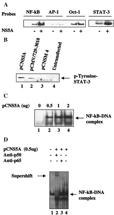
Figure 1.
HCV NS5A activates NF-κB and STAT-3. (A) Activation of cellular transcription factors by NS5A. EMSA was carried out in the presence of 32P-labeled oligonucleotide probes corresponding to NF-κB, AP-1, Oct-1, or STAT-3 in the presence of equal amounts of nuclear lysates prepared from untransfected and Huh-7 cells transfected with wild-type pCNS5A expression vector. (B) NS5A constitutively activates STAT-3 tyrosine phosphorylation. Western blot analysis of STAT-3 protein in cell extracts transfected with wild-type pCMV 729-3010, pCNS5A, or pCNSM4 expression vectors. Cellular lysates were immunoprecipitated with anti-STAT-3 polyclonal serum and Western-blotted with antiphosphotyrosine mAb. Blot was detected by using an ECL kit (Amersham Pharmacia). Lanes 1–3, lysates from cells transfected with pCNS5A, pCMV 729-3010, or pCNSM4 mutant expression vectors, respectively. Lane 4, untransfected lysates. (C) NS5A induces DNA binding activity of NF-κB. EMSA was carried out with 32P-labeled NF-κB probe and increasing concentrations of NS5A-transfected lysates. Lane 1, probe alone; lanes 2–4, NF-κB probe incubated with 0.5, 1, and 2 μg of NS5A transfected lysates, respectively. (D) Supershift of NF-κB protein–DNA complex. EMSA was carried out as described above. Lanes 1 and 4, NF-κB protein–DNA complex; lanes 2 and 3, complex incubated with anti-p50 and anti-p65, respectively.

