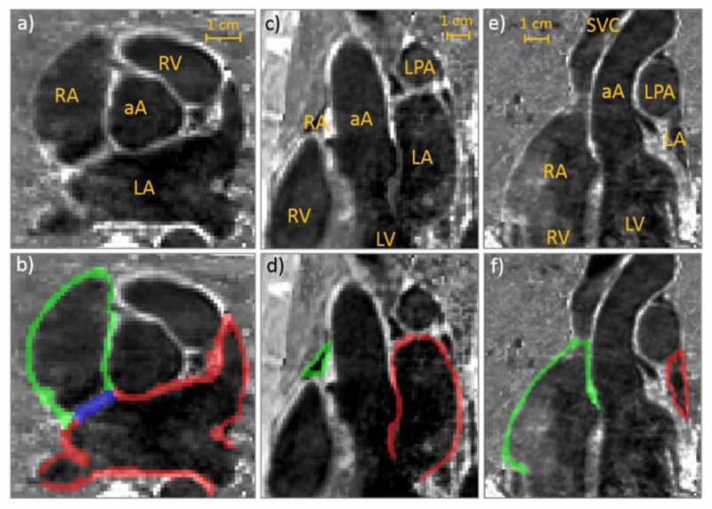
Fig. 3.
a, b) Axial, c, d) sagittal and e, f) coronal views of the atria from one representative subject overlaid, in the bottom row, with the performed manual segmentations. Green: right atrial wall; red: left atrial wall and blue: atrial septum. aA: ascending aorta; LA: left atrium; LPA: left pulmonary artery; LV: left ventricle; RA: right atrium; RV: right ventricle; SVC: superior vena cava.

