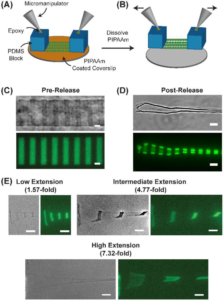
Fig. 2.
Using fiducial marks to measure FN stretch. (A) Schematic of the stretching apparatus where PDMS supports are placed on opposite ends of 1 cm long FN nanofibers. Micromanipulators are then attached to the PDMS supports using high strength epoxy. (B) After hydration and thermally triggered dissolution of PIPAAm, the FN nanofibers release onto the PDMS supports where they are freely suspended in between. (C) Phase contrast and fluorescent image of a FN nanofiber pre-release with 10 µm wide, 10 µm spaced fiducial marks. The center-to-center distance of the fiducial marks was 20.2 ± 0.6 µm. (D) Phase contrast and fluorescent image of a FN nanofiber post-release where the center-to-center distance of the fiducial marks decreased to 10.8 ± 0.4 µm. (E) FN nanofibers were subjected to low, intermediate, and high extensions as indicated by the separation of the fiducial marks. Scale bars are (C and D) 10 µm and (E) 20 µm.

