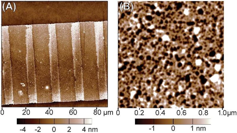
Fig. 3.
AFM images of as-patterned FN nanofibers pre-release while still attached to the PIPAAm-coated coverslip. (A) A large area AFM scan showing the FN nanofiber pre-release with the clearly defined fiducial marks of FN as raised features spaced at regular intervals. (B) A small area AFM scan showing the FN nanofiber molecular morphology in the region between fiducial marks. Note that the FN molecules appear to form an interconnected, fibrillar network with the fibrils forming interconnection points (nodes) where generally 3–5 fibrils meet.

