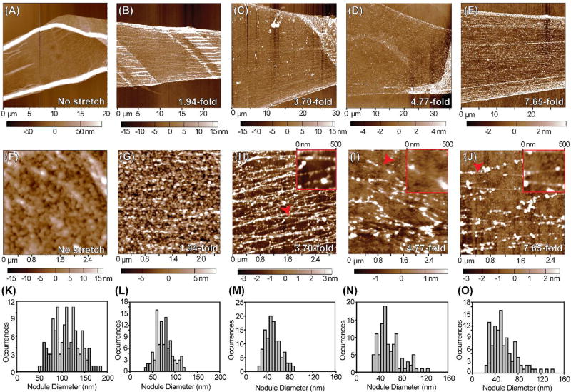
Fig. 4.
Investigating the nanostructure of fully contracted to highly stretched FN nanofibers with AFM. (A–E) Representative low resolution AFM scans of FN nanofibers that were (A) allowed to fully contract in solution prior to imaging, (B) stretched 1.94-fold, (C) stretched 3.70-fold, (D) stretched 4.77-fold, or (E) stretched 7.65-fold relative to the fully contracted state and immobilized prior to imaging. (F–J) Representative high resolution AFM scans of the same FN nanofibers from (A–E). (F) Fully contracted nanofibers had a nanostructure consisting mainly of large, isotropic nodules. (G) FN nanofibers stretched 1.94-fold had an isotropic, nodular network nanostructure with smaller nodules than fully contracted nanofibers. (H) FN nanofibers stretched 3.70-fold had a nodular nanostructure containing FN fibrils that align in the direction of stretch. (I) FN nanofibers stretched 4.77-fold also had a nodular nanostructure containing FN fibrils that align in the direction of stretch. (J) FN nanofibers stretched 7.65-fold had a nanostructure comprised of sparse, aligned FN fibrils that maintain a nodular structure. (K–O) Histograms showing nodule diameters for each amount of extension. (K) Nodules in fully contracted FN nanofibers had a mean diameter of 107 ± 29 nm, consistent with previous findings using smaller FN nanofibers. (L) Nodules in FN nanofibers stretched 1.94-fold had a mean diameter of 73 ± 19 nm. (M) Nodules in FN nanofibers stretched 3.70-fold had a mean diameter of 47 ± 14 nm. (N) Nodules in FN nanofibers stretched 4.77-fold had a mean diameter of 57 ± 20 nm. (O) Nodules in FN nanofibers stretched 7.65-fold had a mean diameter of 53 ± 23 nm.

