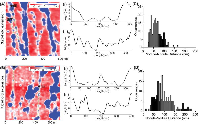
Fig. 7.
Investigating the sub-fibril features of FN nanofibers stretched 3.70-fold and 7.65-fold. (A) A representative region from a 3 µm×3 µm scan of a FN nanofiber stretched 3.70-fold. (A-i and -ii) representative cross-sectional profiles along beads-on-a-string sub-fibril features for 3.70-fold extension. (B) A representative region from a 3 µm×3 µm scan of a FN nanofiber stretched 7.65-fold. (B-i and -ii) representative cross-sectional profiles along beads-on-a-string sub-fibril features for 7.65-fold extension. (C) Histogram showing nodule to nodule distance of FN nodules along beads on a string regions similar to those represented in (A-i and -ii) for 3.70-fold stretched nanofibers. (D) Histogram showing nodule to nodule distance of FN nodules along beads on a string regions similar to those represented in (B-i and -ii) for the 7.65-fold stretched nanofibers.

