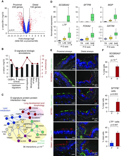
Figure 1.
Distal airway epithelial signature. (A) Volcano plot depicting differential gene expression between the distal (D; n = 63) and proximal (P; total n = 48, including trachea [TR; n = 27] and fourth- to sixth-generation bronchi [n = 21]) small airway epithelium of healthy nonsmokers (SAE-NS). D and P signatures were identified as sets of genes significantly up-regulated (red dots) in corresponding regions. Blue dots represent nonsignificant gene probe sets. (B) Biologic annotation categories enriched in the D signature on the basis of Ingenuity Pathway Analysis (IPA). Shown are the top three IPA-predicted upstream regulators and biologic functions based on the P value (bars, left y-axis) and enrichment score (red boxes, right y-axis). (C) STRING9.1-based protein–protein interaction networks in the D signature. Each circle corresponds to an individual gene; colors distinguish clusters determined using a Markov clustering algorithm and annotated using IPA and/or Gene Annotation Tool to Help Explain Relationships (GATHER). The structure of lines reflects the confidence of association, from thick (most confident) to thin (least confident). (D) Microarray-based normalized expression of selected D-signature genes (see Table E2 for full gene names and Figure E1 for more examples, statistics, and polymerase chain reaction validation) in the epithelium of TR (n = 27), large airway epithelium (LAE; fourth- to sixth-generation bronchi [n = 21]) and SAE (10th- to 12th-generation bronchi [n = 63]) of healthy nonsmokers. The direction of the proximal–distal (P–D) axis is shown. (E) Representative images of the P (bronchus) and D airway samples analyzed using immunofluorescence for D-signature markers secretoglobin family 3A member 2 (SCGB3A2), surfactant protein B (SFTPB), and lactotransferrin (LTF) in combination with cilia markers tubulin β4 chain (TUBB4) or dynein axonemal intermediate chain 1 (DNAI1). Nuclei are stained with 4′,6-diamidino-2-phenylindole (blue); scale bar = 20 μm. See Figure E2 for more examples. (F) Frequency of cells expressing SCGB3A2, SFTPB, and LTF in the P and D airway epithelium (n ≥ 3 samples; total, ≥500 cells/group). BH = Benjamini-Hochberg correction; CEBPA = CCAAT/enhancer-binding protein-α; FOXA2 = forkhead box protein A2; GATA6 = GATA-binding protein 6; MGP = matrix Gla protein; NKX2-1 = NK2 homeobox 1; STK40 = serine/threonine kinase 40.

