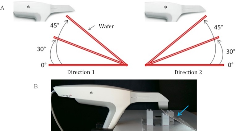
Fig 1.
(A) blueprint of the acquisition parameters for wafer intra-oral scanner imaging. Three angulations: 0°, 30° and 45°. Two directions: “direction 1” on the left; “direction 2” on the right. (B) photo of the setup with the Trios camera (3shape, Copenhagen, Denmark); blue arrow indicates the wafer; asterisk indicates the intra-oral camera.

