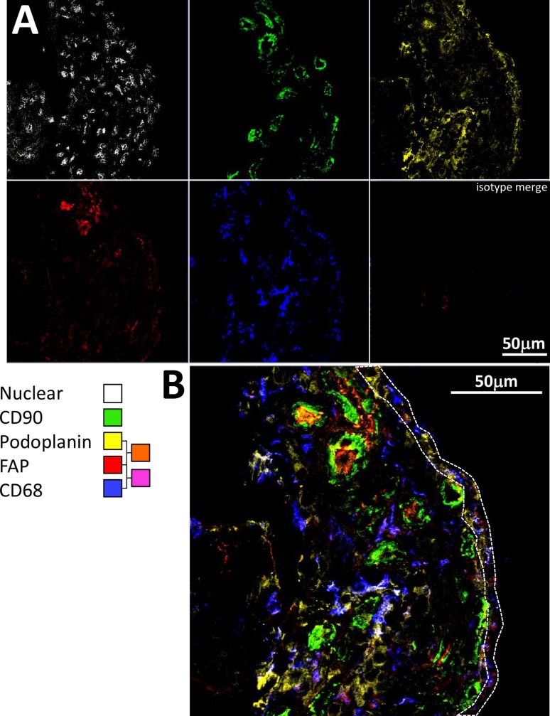
Fig 5. FAP is expressed at low levels in synovial biopsies of patients with self-limiting disease.
(A) Multicolour confocal microscopy images are shown for tissue staining at baseline with FAP (F11-24), podoplanin (D2-40), CD68 (Y1-82A), CD90 (Thy-1A1) antibodies followed by secondary agents, and nuclear (Hoechst) stain in a patient with unclassified arthritis whose disease spontaneously resolved. (B) Higher magnification, merged image. The region representing the lining layer is highlighted by a dotted line.

