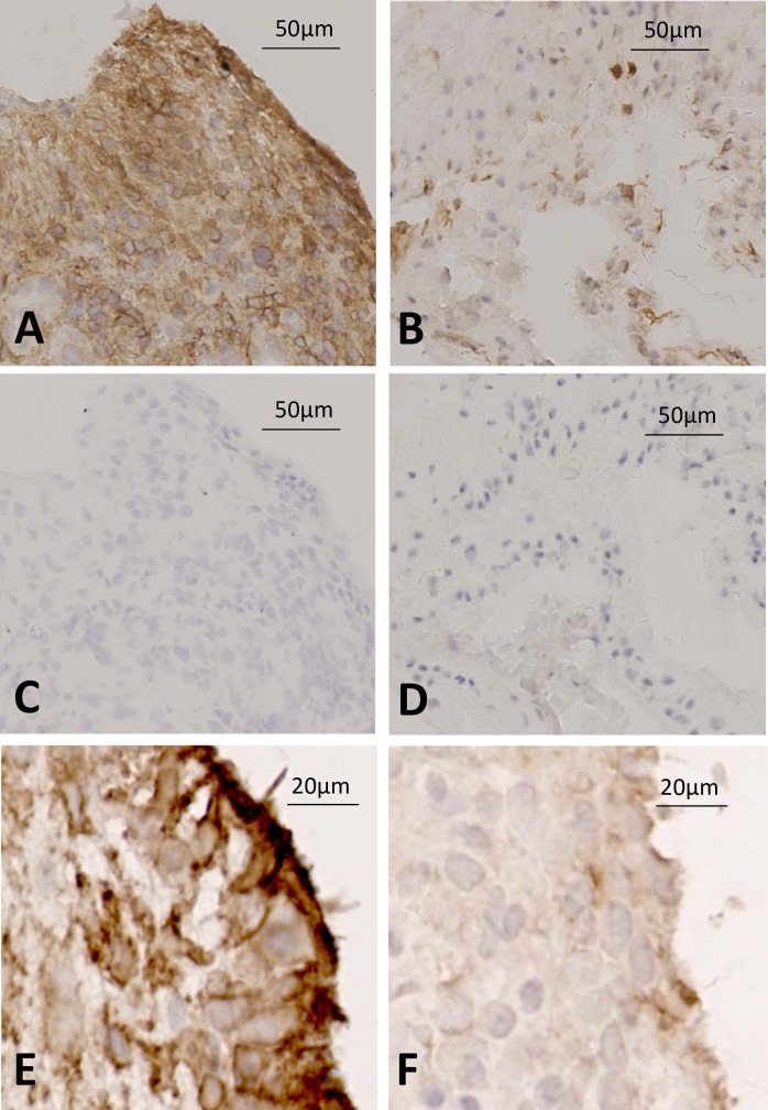
Fig 6. High expression of FAP in tissue from patient developing RA compared to UA control.
Immunohistochemistry was used to stain for FAP positive cells using a sheep anti-FAP antibody in representative tissues from patients developing RA and non-RA disease. (A low power, E high power) FAP staining in patient developing RA vs (C) isotype control; (B low power, F high power) FAP staining in patient with undifferentiated arthritis vs (D) isotype control.

