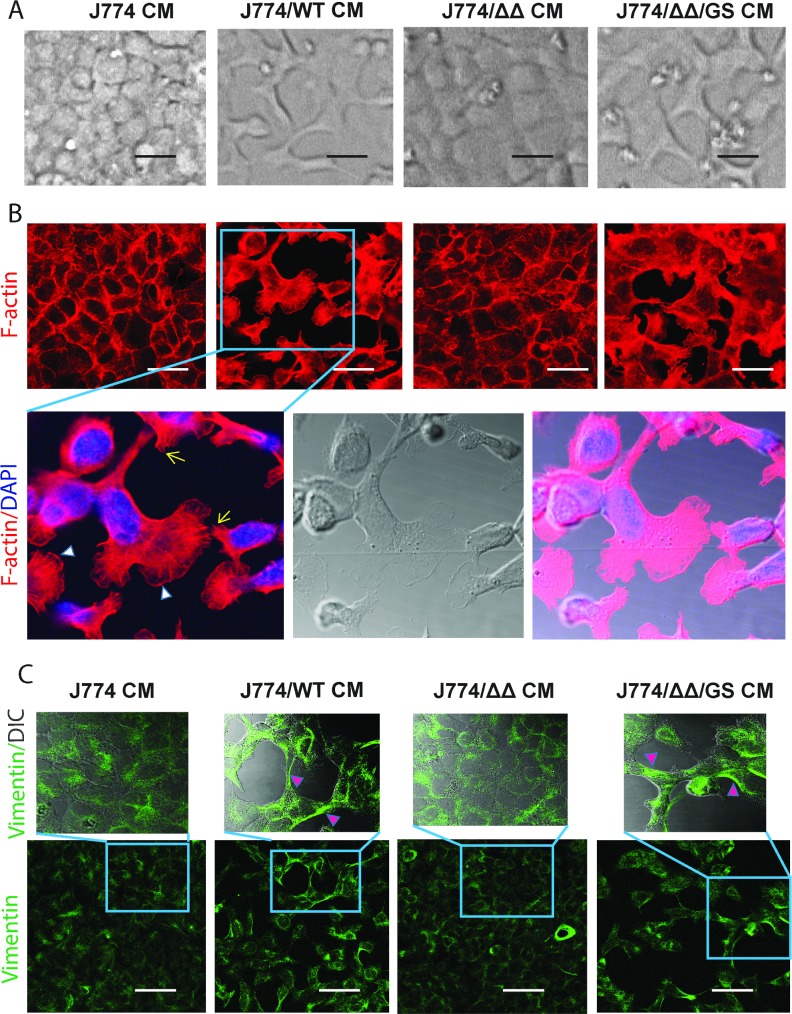
Fig 2. Morphological and cytoskeletal changes in mouse primary epithelial cells (C57BL/6) induced by conditioned media from J774 macrophages co-incubated for 18h with E. faecalis GelE/SprE producing strains.
(A) Phase contrast microscopy images of primary mouse colon epithelial cells incubated with J774/E. faecalis CMs. Representative images of 10 experiments; (B) Immunostaining with rhodamine phalloidin for F-actin (red) and nucleus with DAPI (blue). Lamellipodia and filopodia formation are shown by white arrowheads and yellow arrows respectively; (C) Immunostaining for vimentin. Pink arrowheads indicate intermediate vimentin filaments. Scale bars– 20 μm.

