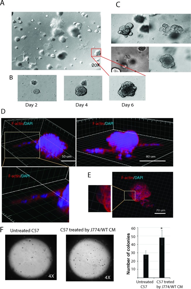
Fig 6. C57/J774/WT cells form distinct type of colonies in matrigel.
(A) Phase contrast images of colonies originated from a single cell suspension of adherent C57/J774/WT cells after 6 days growth in 3D matrigel. (B) Dynamic growth one colony in 3D matrigel by days; (C) Phase contrast images demonstrate different type of colonies as round colonies (upper panel) and colonies with branching (lower panel). (D, E) Reconstructed 3D images of single colonies stained for F-actin (phalloidin, red) and nuclei (DAPI, blue). Images demonstrate (D) cellular extensions and (E) actin-rich cellular protrusions. (F) Epithelial cell suspension in matrigel (50 μl containing ~ 500 cells) was plated into 1 well of an 8-well camber-slide and grown in medium supplemented with 10% FBS. After 1 week of incubation, the colonies were identified. C57cells treated with J774/WT CM give rise to higher number of colonies compared to untreated cells. Statistical analysis was performed using the one-way ANOVA where *P = 0.023 as compared to untreated C57 cells, n = 3.

