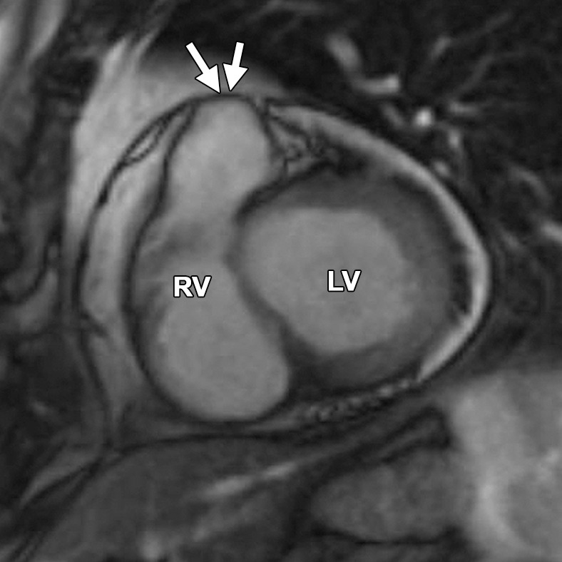
Figure 12d.

Normal pulmonary valve sinuses in a 58-year-old woman. RVOT (a, b) and short-axis (c, d) bright-blood cine SSFP MR images show normal pulmonary valve sinuses at end diastole (a, c) and end systole (b, d). On the short-axis images, the pulmonary valve sinus (arrows in c, d) can give the appearance of dyskinetic systolic wall motion. To avoid misdiagnosis, it is important to identify the level of the pulmonary valve annulus (dotted line in a, b). Normal outpouching of the sinuses (arrow in a, b) occurs above the level of the annulus, whereas wall motion abnormalities in ARVC occur below this level.
