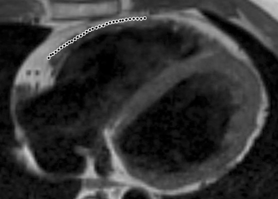
Figure 2b.

Infiltration of fat in the RV of two patients with a history of ARVC. Axial T1-weighted fast spin-echo non–fat-suppressed MR images of a 52-year-old man (a) and a 37-year-old man (b) show fingerlike projections of fat, which has high signal intensity, in the RV wall. Note that the pattern of infiltration of fat resulted in myocardial thinning in a, but myocardial thickness is preserved in b. The dotted line indicates the expected location of the myocardial wall.
