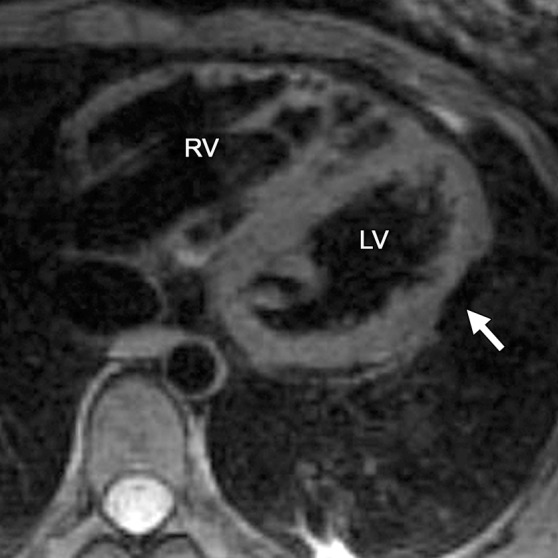
Figure 4b.

Infiltration of fat in the LV of a 30-year-old man with a history of biventricular ARVC. Axial T1-weighted fast spin-echo non–fat suppressed (a) and T2-weighted fat-saturated (b) MR images show the typical infiltration of fat (arrow) in the lateral wall of the LV.
