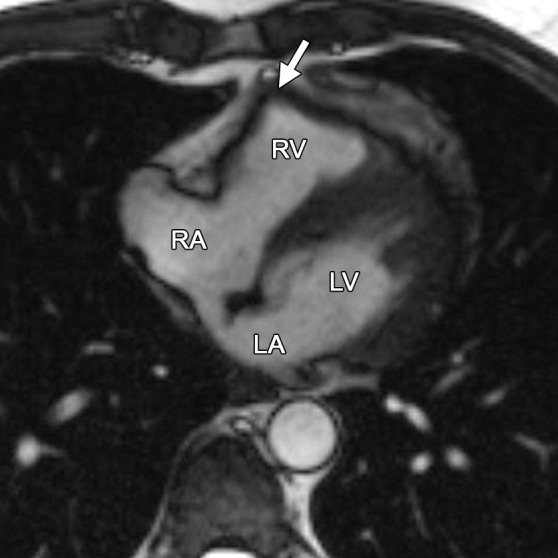
Figure 6b.

RV free wall tether in a 50-year-old man with no history of ARVC. Axial end-diastolic (a) and end-systolic (b) bright-blood SSFP MR images show tethering (arrow) of the pericardium along the RV free wall to the posterior sternum, which may be misinterpreted as dyskinetic RV wall motion. LA = left atrium, RA = right atrium.
