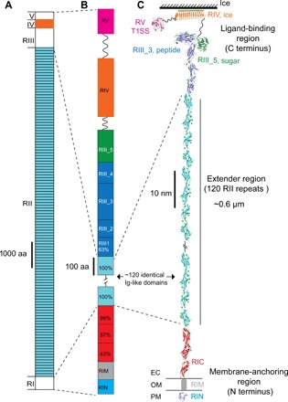
Fig. 1. Overall structure of MpIBP.

(A) Linear domain map of MpIBP drawn to scale. The MpIBP amino acid (aa) sequence is shown in fig. S1. RII and RIV (colored light blue and orange, respectively) are known from two structures solved previously (10, 11, 22). RI, RIII, and RV in white are new three-dimensional structures determined in this study. (B) Expanded view of the RI and RIII to RV linear domain maps colored as in (C). Sequence identities (%) to a 104–amino acid RII repeat are shown for the RIC and RIII_1 domains. (C) NMR and x-ray crystal structures of linked MpIBP domains from N to C termini are shown in cartoon representation: RIN (blue), RIC (red), RII repeats (cyan), RIII_1–4 (dark blue), RIII_5 (dark green), RIV (orange), and RV (magenta). Small green spheres indicate calcium ions. OM is indicated by horizontal lines on either side of RIM. The solution structure of RIM determined by SAXS is illustrated as a gray cylinder. Hatched lines indicate the ~108 RII repeats that are not shown in the figure. The linker regions between RIII_5/RIV (94 residues) and RIV/RV (112 residues) are indicated by wavy lines.
